Heart CT SCAN
What happens during a heart ct scan?
What is a heart CT scan?
A heart scanner is specially designed to study the heart. Most of the time, the aim is to detect partial or total obstruction of the coronary arteries.
An electrocardiogram is used to coordinate the timing of the x-ray pictures between heartbeats, when the heart muscles are relaxed. This allows the technologist to obtain clear images of the heart and coronary arteries.
What is the purpose of a heart CT scan?
A heart CT scan is done to detect obstruction in the coronary arteries. Sometimes the cardiologist orders the scan for other reasons: to measure the thoracic aorta; search for clots in the heart chambers; assess a patient before a procedure such as the insertion of an artificial heart valve; or treat a heart rhythm disorder.
The exam is often performed on patients who meet the following criteria: those with symptoms such as chest pain or shortness of breath; who present risk factors such as high cholesterol, dyslipidemia, hypertension, tobacco use and/or hereditary factors; who have an abnormal coronary calcium score; or to follow up on an equivocal or positive stress test.
What happens during a heart CT scan?
-
1
When you arrive, a receptionist will ask you for your prescription.
In preparation for the exam, we will ask you detailed questions about your medical history to ensure there are no contraindications.
-
2
You will be called from the waiting area and given a small room where you can undress. Then, a technologist will position you on the scanning table and place electrodes on your chest, like for an electrocardiogram
-
3
If approved by the radiologist, the technologist may inject a medication to slow your heart rate if it is too high.
-
4
Before the exam, which lasts around five minutes, you will receive an injection of an iodine-based contrast dye.
-
5
Immediately following the exam, the radiologist will give you the result orally. Before you leave, your images and the exam report will be given to you.
What are the risks of the exam?
A heart CT scan takes pictures using x-rays. Latest-generation scanning devices ensure very low levels of radiation exposure.
The exam requires the injection of an iodine-based contrast agent, which carries a risk of allergy just like any other scan requiring an injection. If you have an allergy, your doctor can prescribe a preventive treatment. If you have already had an allergic reaction to an iodine-based contrast dye but must undergo the exam, bring the report from your previous scan on the day of the procedure so the team can choose a different contrast dye. The allergy is not caused by the iodine itself but by the epitopes contained in the contrast, which vary depending on the product.
Sometimes it is necessary to inject a beta-blocker to slow your heart rate, which may cause your blood pressure to drop. This may be combined with another medication (nitrate derivative) to dilate your arteries.
The technologist will measure your blood pressure right before the exam. In the event of low blood pressure or a contraindication for either of these medications (revealed by your medical history before the exam), they will not be used for the scan. In this case, the scan will not be as efficient, but generally still sufficient to make the diagnosis.
Important: A heart scan is contraindicated if you are pregnant.
How to prepare for a heart scan
On the day of the exam, avoid stimulants such as coffee and tea. We suggest you arrive for your appointment slightly in advance, to avoid extra stress.
You must bring the prescription from your cardiologist or doctor. If a creatinine test (kidney function) was prescribed, you also need to bring the results.
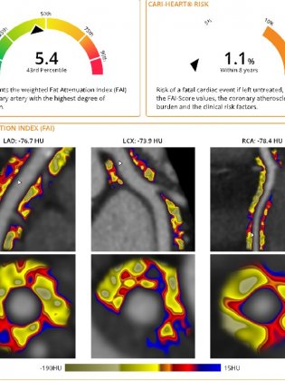
Individualized quantification of the inflammation of the 3 main coronary arteries
An artificial intelligence tool now makes it possible to obtain an individualized quantification of the inflammation of the 3 main coronary arteries.
The CaRi-Heart® software (Caristo company) makes it possible to quantify pericoronal inflammation on a cardiac scanner and to estimate the patient's relative risk of suffering a fatal cardiac event in the next 8 years compared to people of the same age, and of the same sex.
This analysis, offered at the American Hospital of Paris, then allows the doctor to prescribe the most appropriate treatment for the patient in order to reduce risk factors, including inflammation. This analysis can be repeated during subsequent examinations to establish comparisons and follow-up from one examination to another. This follow-up is essential to optimize the care of the patient (hygieno-dietetic rules, treatment with statins). The CaRi-Heart® analysis requires that the anonymized cardiac scans be sent to the Caristo company who will process them and return a detailed report to the doctor.

-
2000
Number of heart CT scans performed at the American Hospital of Paris in 2019. Our imaging center features two scanners, including one ultrafast device more specifically dedicated to heart imaging procedures.
Delivering premium customer service and medical monitoring to patients is a priority at our hospital.
What is calcium score?
Learn more