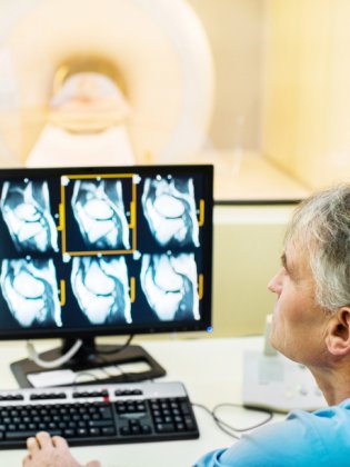
Pediatric MRI of the brain under anesthesia
My child is uncooperative or has autism or symptoms of autism and needs to undergo an mri of the brain under anesthesia. What can we expect during this exam?
An MRI of the brain requires cooperation from the patient, who mustn’t move for the duration of the exam (30 to 40 minutes). This constraint can lead to opportunity loss for many anxious, agitated or uncooperative children and young adults, depriving them of an exam that is essential to their diagnosis, treatment or follow-up care. The American Hospital of Paris is the only hospital to offer this procedure under short-term anesthesia for patients with autism, epilepsy, delayed development and neurodevelopmental disorders.
What is magnetic resonance imaging (MRI)?
Magnetic resonance imaging (MRI) uses a magnetic field (magnet) and radio waves to capture images of the human body thanks to the body’s hydrogen atoms. Placed within a magnetic field, all the hydrogen atoms turn to face the same direction. When stimulated by a radiofrequency pulse, they become “resonant” with the waves. When the pulse stops, the atoms release their stored energy by producing a signal, which is recorded and processed into images by a computer. No ionizing radiation is emitted.
The MRI room is equipped with a machine comprising a tunnel-shaped magnet into which the scanning bed slides, and an antenna adapted to the various regions of the body to be explored.
What is the purpose of an MRI of the brain?
MRI is the most precise imaging technique available for studying the brain. It can detect congenital morphological defects, enabling patients with epilepsy to seek neurosurgical treatment. It can highlight developmental disorders in the brain; assess vascularization and insufficient blood flow to the brain; and depict cerebrospinal fluid circulation in the brain’s ventricles.
In patients with autism, an MRI of the brain can detect possible inflammation, a developmental problem or insufficient blood supply to certain areas of the brain. In many developmental disorders, an MRI of the brain is the first step before conducting more in-depth, particularly genetic, investigations.
Vingt ans de consultations de génétique clinique sur site dans les hôpitaux de jour pour les personnes atteintes de troubles du spectre autistique de la région parisienne
Contraindications, risks and drawbacks of MRI
An MRI is a totally painless exam, but it is long and unpleasant due to the noise produced inside the machine. When under anesthesia, the patient doesn’t hear the noise.
The few contraindications and risks arise mainly from the use of a magnetic field. Any ferromagnetic materials implanted in the body which could be dislodged during the exam and any electromagnetically adjustable materials may present a risk for the patient or cease to function properly after an MRI.
Absolute contraindications for MRI include:
- Patients with a pacemaker (note that MRI-safe pacemakers have been available for several years; however, a consultation with a cardiologist before and after the MRI is necessary).
- Patients with metallic heart valves
- Patients with a brain or spinal cord neurostimulation device or a cochlear implant
Patients with a metallic foreign body must be assured of the absence of risk before undergoing an MRI. They include:
- Patients that may have metal fragments in their eyes: a CT scan will determine the exact position of these foreign bodies in the eye socket.
- The same precautions must be taken for patients with a metallic foreign body near the vascular structures, particularly the arteries: an imaging exam must be done to verify the position of the foreign body in relation to the nearest organ.
As a precaution, best practice dictates that patients who have undergone a surgical implant of ferromagnetic material (artificial joints in particular) or vascular stents should wait at least three weeks after the surgery before undergoing an MRI. It is also important to know the exact nature of the material, in order to determine whether it is MRI-safe.
Allergies to the contrast medium (gadolinium) used for MRI occur far less frequently than allergies to iodine. The observed frequency of allergic reactions to gadolinium in children is from 1 to 4 /10,000; these reactions are almost always benign.
Contraindications and risks associated with anesthesia
At the American Hospital of Paris, the anesthesia protocol for children with autism includes the administration of clonidine, propofol and/or sevoflurane. The side effects and risks of these drugs are mainly respiratory, including hypoventilation, blocked breathing, regurgitation and inhalation of gastric contents (aspiration pneumonia).
Patients must refrain from eating solids for six hours and from drinking clear liquids for one hour before the MRI.
To ensure your child’s safety during the exam, we use airway management devices (laryngeal mask airway or endotracheal tube) and assisted ventilation. The new MRI room at the American Hospital of Paris is equipped with a non-magnetic ventilator. Patients are monitored by the anesthesiologist, who is present in the MRI room for the duration of the exam.
The effects of the anesthesia wear off after 20 to 30 minutes. However, full recovery of the upper body functions may take several hours. After being in the recovery room, your child will continue to be monitored in a resting lounge where he can have something to eat or drink and gradually resume normal activity before going home.
If you plan to return home with your own vehicle, an adult escort must be present in addition to the driver. Once home, your child may feel tired and need to sleep for a few hours, or she might have a temporary burst of energy. Whatever the case may be, there is no need to worry: simply let her resume normal activity at her own pace.
However, if he develops a fever above 38.5° C (101.3° F) or is unable to stay hydrated due to repeated vomiting, you must contact us.
What happens during an MRI of the brain?
When you come to the MRI department, you and your child will be escorted to the pre-procedure room where a nurse will administer the pre-medication. After removing her clothes and any metal objects she is wearing including a watch, jewelry, glasses, and auditory or dental prostheses, she is positioned on the MRI stretcher. She keeps only the tablet or favorite toy you have brought with you.
The nurse inserts the IV line if this is the induction method that was chosen during the anesthesia consultation. If the anesthesia is to be administered by inhalation, we begin by asking the child to breathe with the face mask. The American Hospital of Paris anesthesia team applies the principles of Applied Behavioral Analysis (ABA). Our goal is to bring your child to a state of unconsciousness without disrupting his routine or ever forcing or rushing him. We always ask that a parent, or an educator accustomed to communicating with the child, remain in the pre-procedure room.
When your child loses consciousness, you will leave the pre-procedure room and return to the MRI reception area.
We complete the preparatory tasks (place electrodes and monitoring wires, start perfusion) and take a blood sample for lab tests.
Then your child is taken to the MRI tunnel. The electrodes and monitoring wires are hooked up to the MRI monitor, and the ventilator circuit is connected to the amagnetic ventilator. These steps are monitored by the anesthesiologist, who is present in the MRI room for the duration of the exam. A contrast agent may be injected to improve image quality.
After the MRI, your child, who is still unconscious and lying on the stretcher, is taken to the recovery room. As soon as he wakes up, you will be called in to be with him. The electrodes, monitoring wires and IV line must remain in place until he is transferred to the resting lounge. We will return his tablet or favorite toy to him. He will be allowed to drink one hour after the end of the exam and to eat shortly thereafter if the wake-up continues to go smoothly.
Once home, having bravely endured the MRI procedure under anesthesia, your child may feel tired and need to sleep for a few hours, or she might have a temporary burst of energy. Whatever the case may be, there is no need to worry: simply let her resume normal activity at her own pace. However, if he develops a fever above 38.5° C (101.3° F) or is unable to stay hydrated due to repeated vomiting, you must contact us.

How to prepare for an MRI
The day of the procedure:
Go to the MRI department on Level -1. Your child must not eat any solids for six hours and must not drink clear liquids for one hour before the procedure. He must not be carrying or wearing any metallic objects such as a watch, ring, earrings or belt. Medications containing methylphenidate such as Ritaline®, Quasym® or Concerta® must be discontinued the day before the MRI.
You must bring:
- Carte Vitale insurance card
- MRI prescription
- Reports from any previous MRIs
- Consent form signed by both parents
- Tablet or favorite toy
Anesthesia consultation (around one week before the MRI):
Go to the Pediatric Department on the ground floor of the hospital.
You must bring:
- Health record (carnet de santé)
- Letter from the child’s care facility detailing current care and medications
- Any hospitalization or surgery reports
At this time you may also finalize remaining any paperwork.
Key figures
42 percent of the time, an MRI of the brain detects defects that are non-specific but manifest in young persons with autism.
For patients with epilepsy, an MRI raises the detection level of surgically curable focal brain lesions from 20 percent to 80 percent.
Today, hundreds of children and young adults do not undergo MRI exams because the appropriate sedation/anesthesia is unavailable. This is an obvious loss of opportunity for them.
The American Hospital of Paris is the only hospital to offer MRI under anesthesia for patients with autism, epilepsy, delayed development and neurodevelopmental disorders.
Request an appointment if you already have a prescription
Learn moreRequest an appointment with our geneticist
Learn more