Shoulder Osteoarthritis
L’omarthrose signifie littéralement "arthrose de l’épaule
The role of this cartilage is to ensure that the joint surfaces glide painlessly against one another to enable motion in the shoulder. Cartilage breakdown causes bones to rub against each other, resulting in pain. It can also lead to bony outgrowths called osteophytes, or bone spurs.
Shoulder arthritis is debilitating because it strongly affects a person’s ability to use their arms. The condition generally occurs more frequently in women. Sometimes, both shoulders are affected.
Two types of shoulder osteoarthritis
-
1
Primary shoulder osteoarthritis
Primary shoulder osteoarthritis results from damage to the cartilage and only accounts for three percent of all cases of shoulder osteoarthritis. It can result from an inflammatory disease such as rheumatoid polyarthritis, or from another condition such as osteochondromatosis, characterized by small cartilage tumors: “Imagine the joint like a car wheel: the tire wears down little by little and eventually you’re riding on the rims. That’s when the pain starts.”
-
2
Secondary shoulder osteoarthritis
Secondary shoulder osteoarthritis results from rupture of the shoulder tendons. The shoulder has a group of muscles and tendons called the rotator cuff. The rotator cuff has four tendons (one in front of the shoulder joint, one on top and two at the rear) whose role is to keep the shoulder joint stable while allowing it to rotate. Frequently, one of these tendons ruptures. Following the rupture, the “ball” part of the shoulder’s ball-and-socket joint gradually slides upward into a position where it should not be. This causes rubbing, which leads to osteoarthritis.
What are the symptoms of shoulder osteoarthritis?
The main symptom of shoulder osteoarthritis is pain.
The pain can be described as diffuse, meaning it is felt in the arm and back of the neck rather than in one precise location. This pain, triggered by movement of the shoulder, causes the joint to become increasingly stiff. The patient progressively loses mobility, and it becomes difficult, or even impossible, to raise the arm or reach behind the back. The condition can also lead to debilitating pain at night, causing the patient to wake up frequently.
Wearing of the cartilage is a gradual development that does not occur overnight. At first, shoulder osteoarthritis symptoms are not easy to identify. The condition is relatively well-tolerated by patients at the beginning, when inflammation causes only intermittent pain. The patient may also experience temporary blockage of the shoulder and a “grinding” sensation.
In general, as the arthritis evolves, the pain becomes daily. This can result in the patient avoiding all shoulder movement, causing complete loss of function.
When should I see a doctor for shoulder osteoarthritis?
To avoid the risk of daily pain and total loss of function, patients should see their physician as soon as the first symptoms appear. The patient can begin with a visit to his or her primary physician or a rheumatologist, who will be able to diagnose the osteoarthritis and prescribe the appropriate treatment. If surgery is required, the patient should see an orthopedic surgeon specializing in the shoulder.
What tests are used to diagnose shoulder osteoarthritis?
The specialist starts by doing a clinical exam. He or she will evaluate the patient’s symptoms and level of pain. Several symptoms indicate potential shoulder osteoarthritis.
Following the clinical exam, other tests are done to confirm the diagnosis, such as standard front-to-back and lateral X-rays to visualize the shoulder bones. The radiologist will look for several signs, including an abnormally thin joint, a humerus head that has lost its ball-like shape, or the presence of bone spurs. The images will also help determine whether the patient has primary or secondary osteoarthritis, as well as its stage: early, mild or severe.
In addition to the X-rays, an ultrasound might also be done, allowing the physician to assess the tendons in the shoulder.
Lastly, a CT scan helps to evaluate the state of the rotator cuff and the bones, before possible replacement surgery. The goal of these complementary tests is to determine the best treatment for the patient.
What are the treatments for shoulder osteoarthritis?
The doctor will generally begin with medication and physiotherapy, sometimes combined with intra-articular injections. Physiotherapy is helpful in maintaining whatever mobility remains.
Some doctors use platelet-rich plasma therapy, or PRP, which consists in injecting platelets from the patient’s own blood into the painful joint. This concentrate of platelets is rich in growth factors and helps regenerate damaged cartilage. However, the method's efficacy has not yet been proven. Over time, this type of treatment becomes less effective. When that happens, surgery is sometimes necessary.
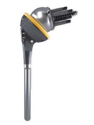
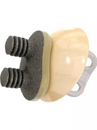
In this case, shoulder replacement surgery with a prosthetic implant may be proposed.
This type of surgery may be necessary when the patient suffers from: osteoarthritis, which causes severe cartilage wear; a complex shoulder fracture; or debilitating and unrepairable lesions in the rotator cuff tendons. There are two types of prosthetic shoulder implants: reverse and anatomic. Mechanically speaking, these solutions are different from each other and are not indicated under the same conditions.
The American Hospital of Paris benefits from the latest technological advances in shoulder replacement surgery techniques, including the use of mixed reality to optimize the implant insertion procedure.
Reverse total shoulder replacement
This procedure is called “reverse” because the surgeon reverses the natural position of the shoulder’s ball-and-socket joint, attaching the ball-shaped part of the implant to the shoulder blade, and the socket to the upper arm bone. This technique enables the shoulder to function even if the rotator cuff is severely damaged. By lowering the humerus bone and restoring function to the deltoid (shoulder) muscle, the lack of function of the rotator cuff tendons is compensated.
The reverse shoulder replacement procedure lasts around 45 minutes and is performed by an orthopedic surgeon specialized in the shoulder. It is generally performed on an outpatient basis (with no overnight hospital stay), but sometimes requires one night of hospitalization.
At the beginning of the procedure, the anesthesiologist numbs the shoulder before administering a light general anesthesia. The operation leaves a scar measuring approximately 10 cm. Rehabilitation can be done through a private practice or hospital, and only in certain cases is a stay in a rehabilitation facility required.
After the operation, a sling is worn at night and during part of the day to immobilize the arm. Use of the arm is restricted to simple daily activities. Rehabilitation begins the very next day after surgery, with physiotherapy as well as exercises done by the patient at home. The surgeon will indicate the frequency and duration of rehabilitation as well as the steps for regaining mobility in the arm.

Anatomic total shoulder replacement
When the rotator cuff tendons are functional, the anatomic shoulder prosthesis reconstructs the shoulder as it was before it became impaired, using personalized implants that imitate the shoulder joint’s natural ball-and-socket anatomy.
Anatomic total shoulder replacement surgery is generally performed on an outpatient basis, but in some cases it may require two nights at the hospital. At the beginning of the procedure, the anesthesiologist numbs the shoulder to ensure greater postoperative comfort, then administers a light general anesthesia. The surgeon then makes a 10 cm-long incision to insert the prosthetic implant.
After the operation, the arm is immobilized with a sling day and night for several weeks. Rehabilitation begins the very next day after surgery. In addition to exercises the patient does at home, rehabilitation with a physiotherapist can be done either through a private practice or at the hospital, but does not necessarily require staying in a postoperative rehabilitation facility.

Book an appointment
Learn more